Dorsal funiculus: All you need to know about nerve fibers enclosed by the perineurium
A funiculus is a little heap of axons (nerve filaments), encased by the perineurium. A little nerve might comprise of a solitary funiculus, however, a bigger nerve will have a few funiculi gathered together into bigger packs known as fascicles. Fascicles are bound together in a typical film, the epineurium.
Physical classification for a cordlike construction or part, particularly one of the huge heap of nerve plots that make up the white matter of the spinal rope. (adj. adj funicular. )
Front funiculus ventral funiculus.
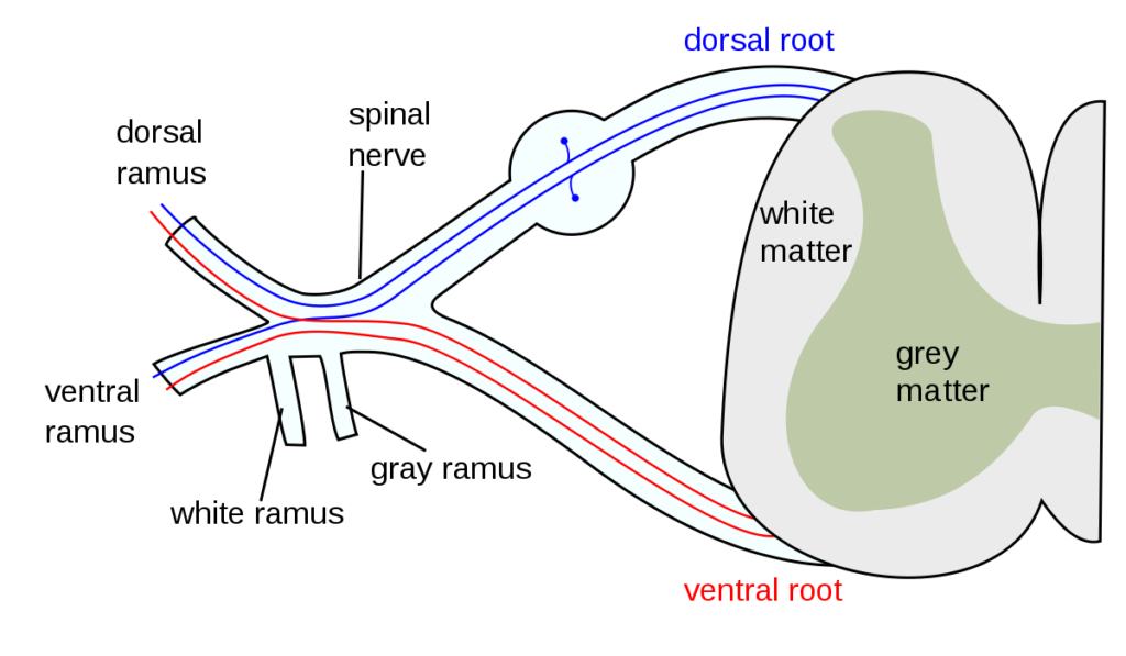
Dorsal funiculus is the white substance of the spinal string lying on one or the other side between the back middle sulcus and the dorsal root.
Horizontal (funiculus latera’lis) is the sidelong mass of strands on one or the other side of the spinal line, between the anterolateral and posterolateral sulci.
Back funiculus dorsal funiculus.
Funiculus sperma’ticus the spermatic rope
Ventral funiculus is the white substance of the spinal rope lying on one or the other side between the ventral middle gap and the ventral underlying foundations of the spinal nerves.

The white matter of the spinal rope
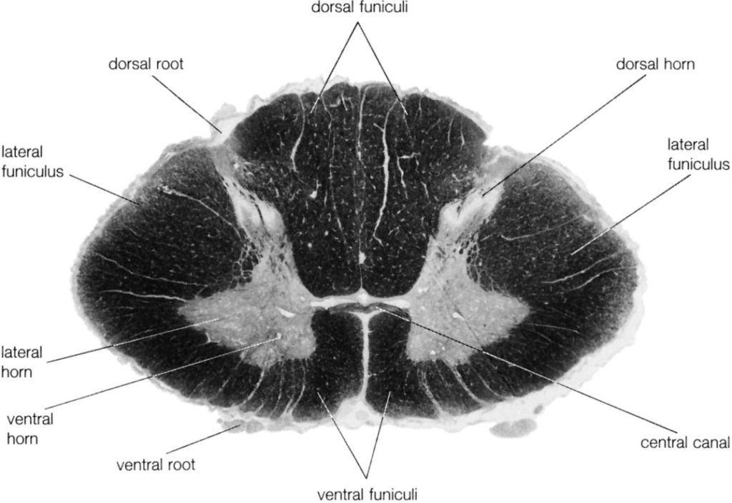
A layer of white matter encompasses the dark matter except for where the dorsal horn contacts the edge of the spinal string. The white matter comprises generally longitudinally running axons and glial cells. An enormous gathering of axons that are situated in a given region is known as a funiculus (for example back funiculus). More modest heaps of axons, which share normal components inside a funiculus are called fasciculus (for example fasciculus gracilis).
Lots and pathways are terms applied to nerve fiber packages that have a practical undertone. A lot is a gathering of nerve strands with a similar beginning, course end, and capacity (for example spinothalamic lot). A pathway is a gathering of lots with a connected capacity (for example postsynaptic dorsal section pathway).
The horns of dim matter separate the white matter into three segments (funiculi): dorsal, sidelong, and ventral. The limit between the horizontal and ventral sections isn’t particular. It is by and large taken to be in accordance with the arising axons of the most horizontal motoneurons. The two dorsal sections are situated between the two dorsal horns of dark matter and untruth next to each other with a typical average line.
The two ventral sections are isolated by a profound crevice, the ventral middle gap, which expands nearly to the extent of the commissural dark matter. Veins travel through the profound crevice as the method of arriving at the focal point of the dark matter.
At the dorsal furthest reaches of the ventral middle crevice is a band of white matter (the ventral white commissure) which associates the two ventral sections.
At the district where the dorsal horn comes to the pial surface of the spinal line, there is a noticeable band of strands called the dorsolateral fasciculus (the plot of Lissauer). This plot contains essential afferent strands, which either rise or slip for a couple of sections prior to entering the dorsal horn.
The dorsal section of white matter is predominantly comprised of the focal cycles of dorsal root ganglion cells. These huge myelinated axons structure the primary pathway passing on skin sensation and position sense (proprioception) from the appendages and trunk to the mind.
Since more filaments are added at each fragment in a caudorostral request, the dorsal segment is tiny in sacral portions and the biggest in the rostral cervical spinal rope. As the filaments from one fragment enter the dorsal segment, they move to shape a strip as near the average edge as could really be expected.
Filaments from the following section move as far medially as they can and structure a strip horizontal to those of the more caudal fragment. When seen at high cervical levels, the filaments from the lower thoracic, lumbar, and sacral sections (fragments beneath T6) structure a particular average strip, the gracile fasciculus.
The more sidelong gathering is wedge-formed and is known as the cuneate fasciculus. It fundamentally contains afferents from the upper thoracic and cervical fragments.
In rodents and numerous non-primate warm-blooded animals, the ventral-most piece of the dorsal segment contains a little however exceptionally unmistakable parcel comprised of little measurement strands which can be effectively recognized from the cuneate and gracile fasciculi.
This gathering establishes the dorsal corticospinal parcel. Since it contributes filaments to each portion as it slides, this plot reduces in size from rostral to caudal levels. The dorsal corticospinal plot strands end in the dorsal horn, halfway dim matter, and, less significantly, on the interneuron pools of the ventral horn.
In primates, carnivores, and some different vertebrates, there is no significant dorsal corticospinal parcel, on the grounds that practically all of the corticospinal strands are found in an unmistakable lot in the dorsal piece of the sidelong segment, a more modest plot in the ventral segment. The sidelong corticospinal plot of primates is surprising in that a portion of its strands make direct synaptic associations with motoneurons in lamina 9. In people, around 20% of the parallel corticospinal filaments make direct corticomotoneuronal associations – by a wide margin the biggest rate in any primate.
The parallel and foremost sections contain an assortment of rising and plunging fiber gatherings. The climbing lots incorporate the spinothalamic, spinocerebellar, and spinotectal plots and the plunging lots incorporate the corticospinal, vestibulospinal, tectospinal, and reticulospinal lots.
Just as these long climbing and dropping plots, there are numerous filaments in the white sections that interface one spinal line portion with another. These filaments are called propriospinal because they regularly lie exceptionally near the dim matter. The biggest propriospinal pathways interface the brachial and lumbosacral amplifications to arrange appendage developments.
White Matter
The white matter of the spinal string is partitioned into three huge areas, every one of which is made out of individual parcels or fasciculi. The back funiculus is situated between the back middle septum and the average edge of the horn. At cervical levels, this region comprises the gracile and cuneate fasciculi; altogether, these are normally alluded to as the back (dorsal) sections.
The sidelong funiculus is the space of white matter situated between the posterolateral and anterolateral sulci. This district of the rope contains clinically significant rising and sliding lots. Those generally significant in the conclusion of the neurologically debilitated patient are the horizontal corticospinal plot and the anterolateral framework (ALS).
Situated between the anterolateral sulcus and the ventral (front) middle gap is a relatively little area, the foremost funiculus. This region contains reticulospinal and vestibulospinal filaments, parts of the ALS, the foremost corticospinal plot, and a composite group called the average longitudinal fasciculus (MLF).
Two little yet significant parts of the white matter are the front white commissure and the posterolateral (dorsolateral) lot. The foremost white commissure is situated on the front midline and is isolated from the focal trench by a thin band of little cells. The posterolateral parcel, the lot of Lissauer, is a little heap of softly myelinated and unmyelinated filaments covering the back horn.


